
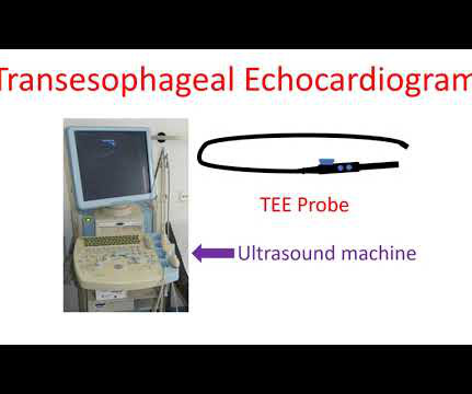
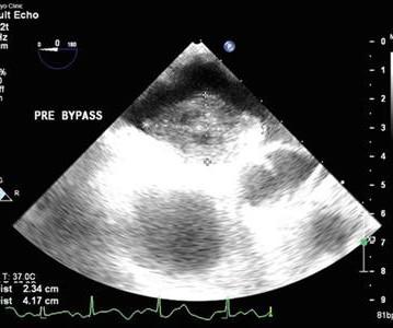
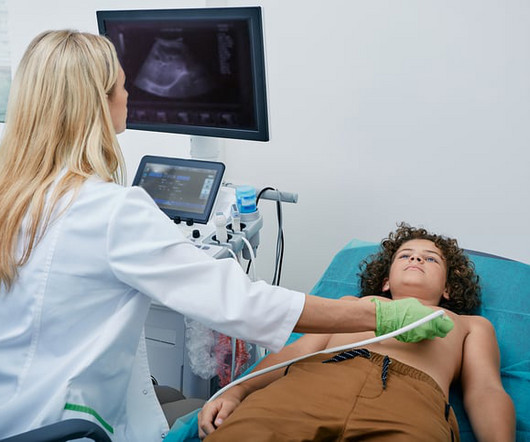
Transesophageal echocardiogram
All About Cardiovascular System and Disorders
MAY 7, 2024
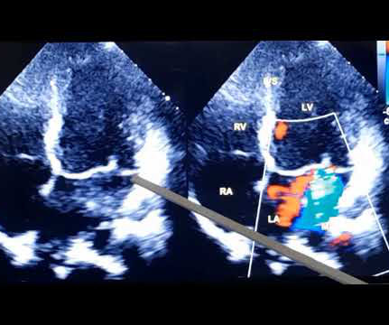
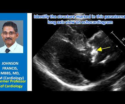
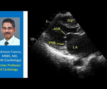
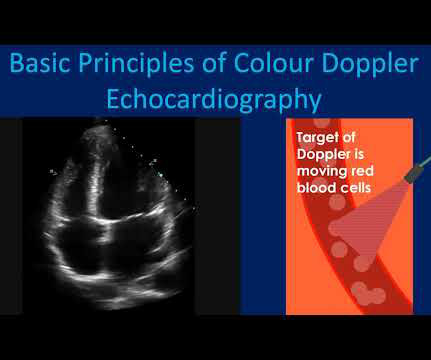
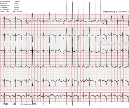
Echocardiogram is an image of the heart using ultrasound. Transesophageal echocardiogram or TEE test, is obtained by introducing a special type of transducer, also called a TEE probe, through the throat into the food pipe (esophagus) and stomach. Usual echocardiogram is obtained by placing the transducer or probe on the chest.


Let's personalize your content